AATVar : a database of human alpha-1 antitrypsin variants
Mprocida
Mutation sequence analysis
Contributed by
CHU Lille
HGVS nomenclature (NM_000295.4)
Usual nomenclature (Without signal peptide)
Nomenclarure including the signal peptide
c.194T>C
Type of variation
AAT variant
Mutation Location
Exon 2
Genetic background
ACMG classification
Pathogenic
Comments
rs28931569
AAT variant and Q0 alleles
Variant name
Mprocida
Also Known as
pathogenicity
Deficient
HGVS nomenclature protéine
p.Leu65Pro
3D position of aa affecteded
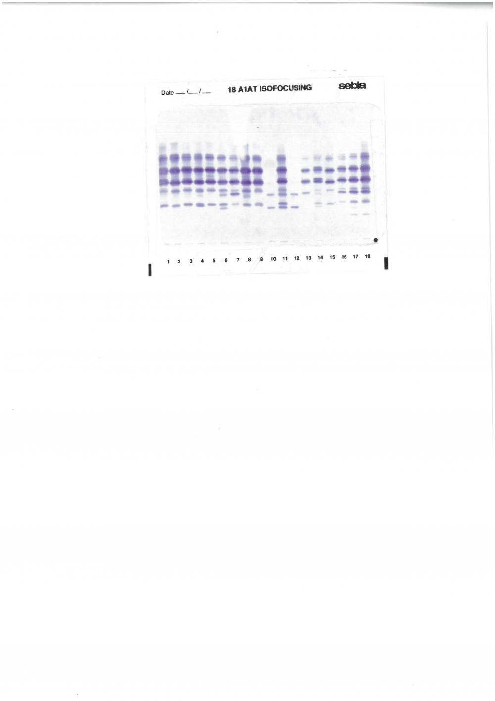
Mobility on polyacrylamide gel
Mobility on agarose gel
M
AATserum level (g/L)
Heterozygous
0.71
Homozygous
Anti-elastolytic activity (IU/L)
Heterozygous
9963
Homozygous
Comments
Associated with a M2 allele.
Evaluation of the crystallographic structure of alAT suggests the "Leu to Pro“ mutation may disrupt α-helix A in the region of Pro21-Ser45,suggesting the possibility that the a1AT Mprocida molecule is unstable and degraded intracellularly prior to secretion. No liver accumulation.
The Leu-Pro substitution involves two uncharged amino acids, a fact that is consistent
with the observation of a very small difference in electrophoretic mobility between Mprocida and the common normal M1(Val213)
Occurrence
Ethnic background without frequency range
Ethnic background and frequency
Frequency range
from (%)
0.01
To (%)
0.03
Group tested
Size
Description (who was tested)
Occurrence comments
From gnomAD
Overall comments
Occurrence comments
This variant was identified at a heterozygous status in a 22-year old man presenting with alpha-1 antitrypsin deficiency. It was also identified at a heterozygous status with a M1 variant in a 43-year old man presenting with COPD and in a 75-year old man presenting with pulmonary emphysema and with a I variant in a 66-year old man presenting with pulmonary emphysema.
Evaluation of the crystallographic structure of alAT suggests the "Leu to Pro“ mutation may disrupt α-helix A in the region of Pro21-Ser45,suggesting the possibility that the a1AT Mprocida molecule is unstable and degraded intracellularly prior to secretion. No liver accumulation.